Two column articular distal radius fracture 2R3C3.1
Score and Comment on this Case
Clinical Details
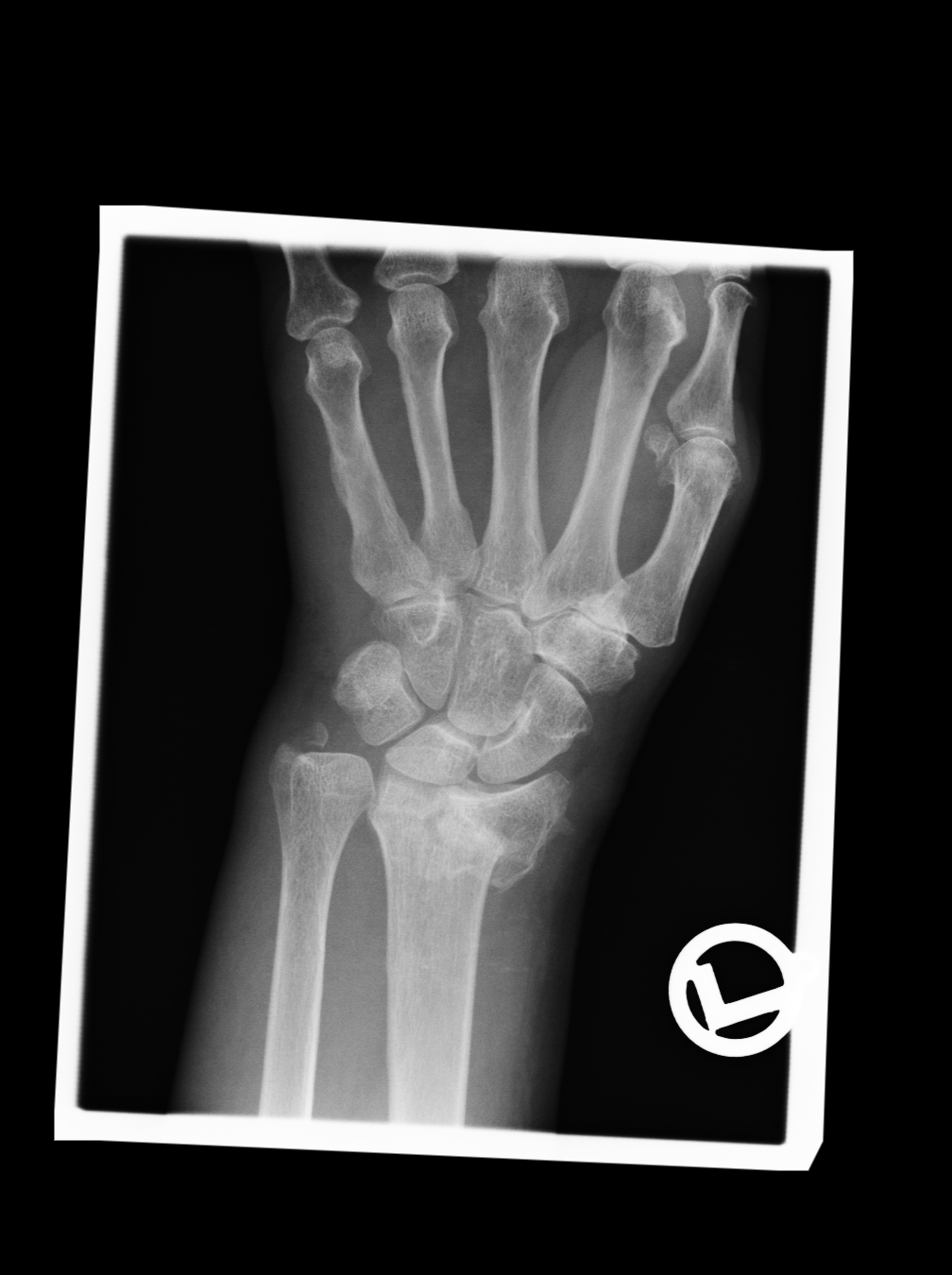
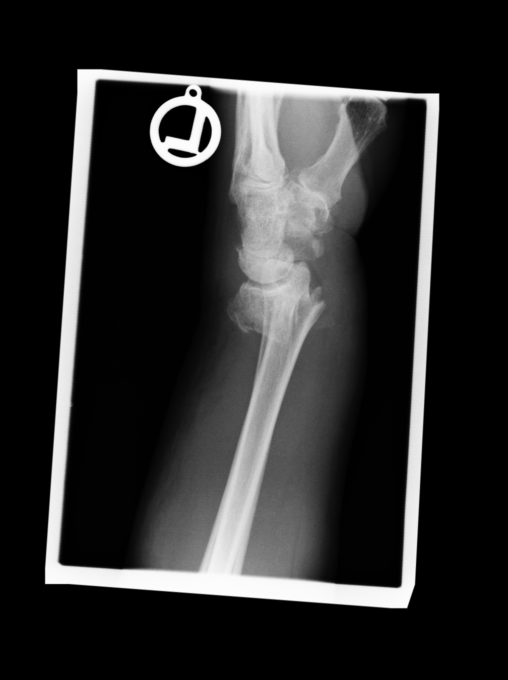
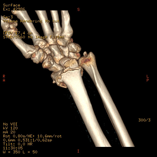
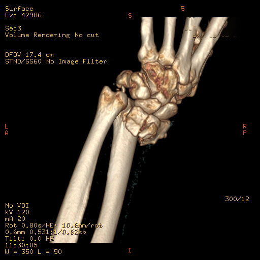
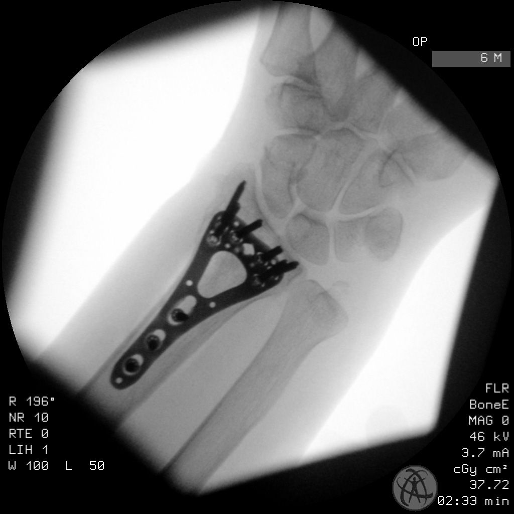
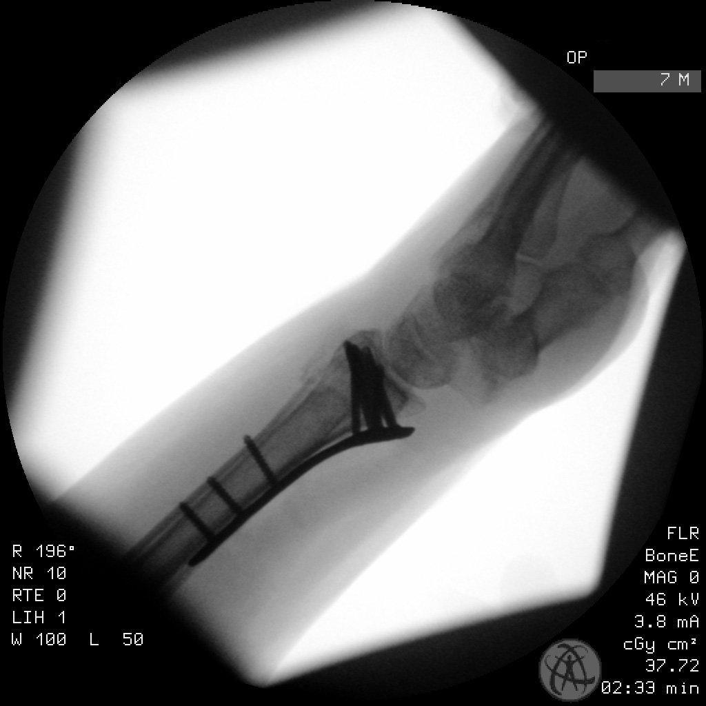
Clinical and radiological findings: This distal radius fracture belongs to a 67-year-old female following a simple trip and fall. On initial plain film imaging (at least based on the lateral projection) you could be convinced that this is a normal dorsally displaced Colle's type fracture. However careful evaluation of the AP film demonstrates a discontinuity between the scaphoid and lunate facets. The exact degree of comminution of the articular surface however was not fully appreciated until completion of a CT scan as part of our standard preoperative planning. Here on the two 3D reconstructions provided one can appreciate a) in the palmar view a complete dissociation between the two joints facets and b) how the lunate facet has a secondary coronal split dividing it into a palmar and dorsal components. The operation took place supine with the arm prepared on a secondary arm table to the side of the main operating table. A standard Henry’s approach over FCR was utilized. I reconstructed both the radial scaphoid facet and the ulnar lunate facet as two individual blocks before reducing them together to the proximal radial metaphysis. This reduction was held with multiple K wires before placing a two column distal radial locking plate. You can see by screw placement in the locking plate that each facet block was fixed individually - each with three locking screws. The middle screw hole was not utilized as this sat directly in the fracture zone between both fragment constellations.
Preoperative Plan
Planning remarks:
Surgical Discussion
Operative remarks:I quite like this case not only due to the good result in reconstructing the articular surface but how it elegantly demonstrates the 2 column nature of intra-articular radius fracture pathology with descrete involvement of the individual facets of the articular surface. This is then reflected in the final placement of the locking screws in the plate - quite literally utilising the 2 columns of the 2 column plate to their fullest extent.
Orthopaedic implants used: DePuy Synthes Variable Angle LCP Two-Column Volar Distal Radius Plate 2.4
Search for Related Literature

Dr Ed Oates
- Germany , Schleswig Holstein
- Area of Specialty - General Trauma
- Position - Specialist Consultant

Industry Sponsership
contact us for advertising opportunities
Article viewed 1404 times
15 Mar 2021
Add to Bookmarks
Full Citation
Cite this article:
Oates, E.J. (2021). Two column articular distal radius fracture 2R3C3.1. Journal of Orthopaedic Surgery and Traumatology. Case Report 18327774 Published Online Mar 15 2021.