Condylar split - a comminuted articular distal humerus fracture 13C3.3
Score and Comment on this Case
Clinical Details
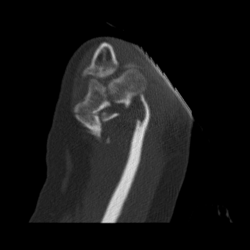
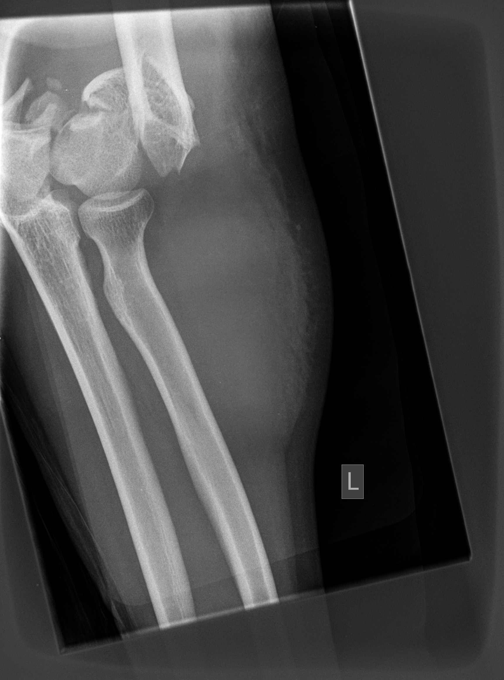
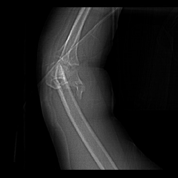
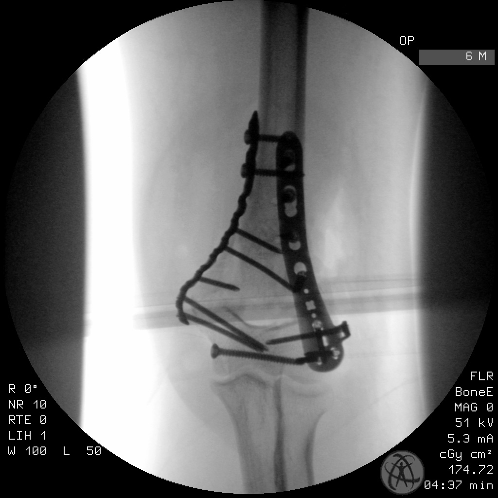
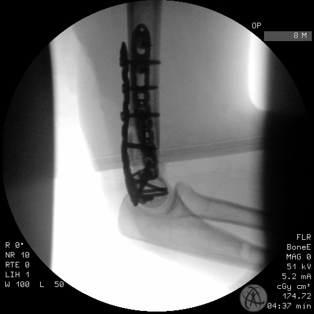
Clinical and radiological findings: This articular condylar distal humerus fracture belongs to an 18-year-old female involved in a motor vehicle accident. A closed a neurovascular intact injury, the fracture/joint was initially immobilized with an Ex-Fix pending definitive operative management in the following days. The pre-operative imaging was restricted to the limited reconstructions of the CT trauma scan and also a single plane film radiograph of the joint in which the fracture ist not completely imaged. One can nonetheless identify a displaced comminuted intra-articular condylar fracture of the distal humerus. The capitellum and trochlea components of the condyles have been split sagittally. There is gross dorsal displacement of the fracture components. Following consolidation of the soft tissue we proceeded with a definitive fixation of the fracture several days later. The operation was performed prone with the arm hanging over a small attachment to the operating table allowing elbow flexion over 90°. A simple dorsal midline approach on the distal arm curving radially around to the olecranon was utilized. Wanting to avoid an olecranon osteotomy we mobilized windows medially and laterally and following neurollysis and mobilsation of the ulnar nerve we achieved a relatively good view across the dorsal aspect of the joint. As the articular surface was predominantly split into two large pieces it was possible, despite the overlying olecranon, to anatomically reduce this joint block and adapt the two components with a single transverse ulnar-to-radial 3.5 mm small frag screw. Following reconstruction of this joint block we reduced this component back to the distal humerus and temporarily held the reduction with multiple K wires. Placement of the dorsoradial and medioulnar plates in 90° plating technique was unremarkable. We achieved good stabilization of the jointblock fragments allowing a stable range of motion on the table. Postoperative protected ROM exercises in a EpicoRom brace was allowed with increasing AROM over 6 to 8 weeks.
Preoperative Plan
Planning remarks:
Surgical Discussion
Operative remarks:I think this is simply a nice case demonstrating what can be achieved without having to osteotomize the olecranon. Reduction of the joint block was tricky with the overlying articular surface, however it was possible. Granted this was not a particularly comminuted/multi fragmentary fracture, and the joints block fragments were large. With more comminution or a more difficult reduction an osteotomy would have been necessary.
Orthopaedic implants used: DePuy Synthes VA-LCP Distal Humerus Plates 2.7/35
Author's Resources & References
Search for Related Literature

Dr Ed Oates
- Germany , Schleswig Holstein
- Area of Specialty - General Trauma
- Position - Specialist Consultant

Industry Sponsership
contact us for advertising opportunities
Article viewed 767 times
15 Mar 2021
Add to Bookmarks
Full Citation
Cite this article:
Oates, E.J. (2021). Condylar split - a comminuted articular distal humerus fracture 13C3.3. Journal of Orthopaedic Surgery and Traumatology. Case Report 18583875 Published Online Mar 15 2021.