Fracture hemiarthroplasty distal humerus
Score and Comment on this Case
Clinical Details
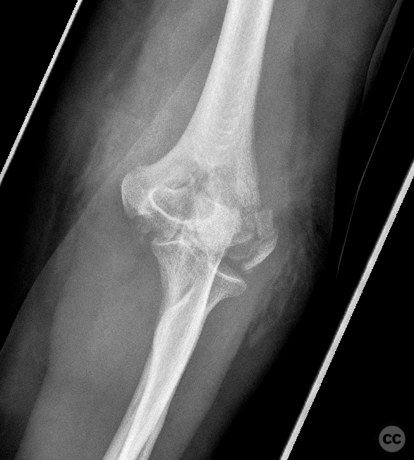
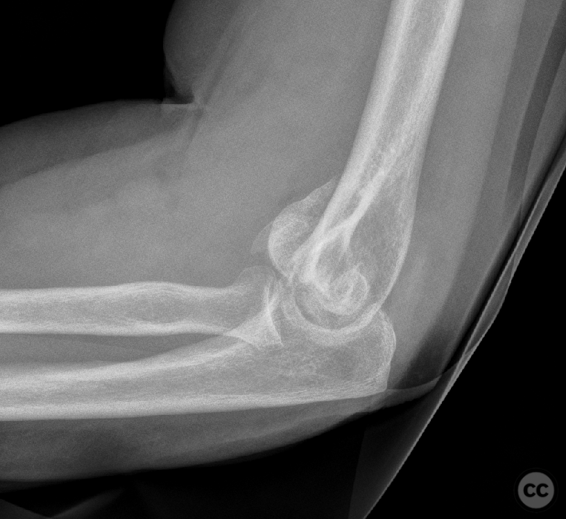
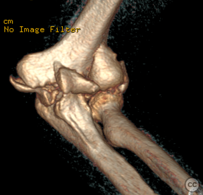
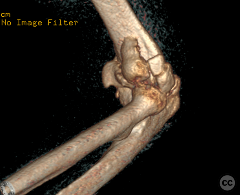
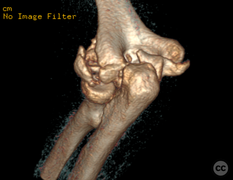
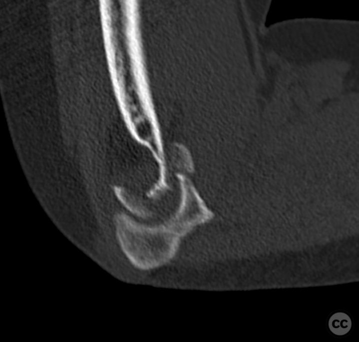
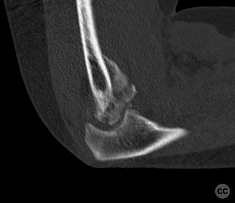
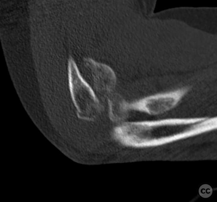
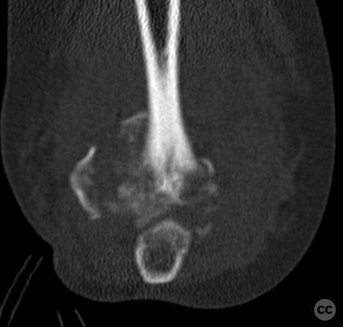
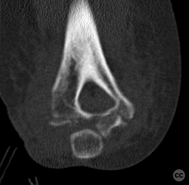
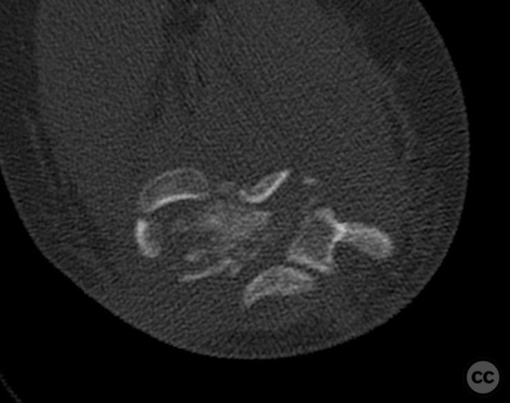
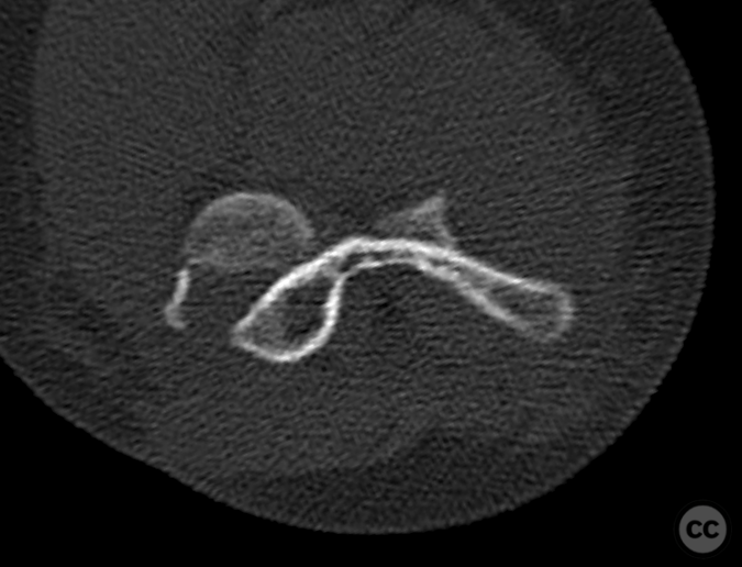
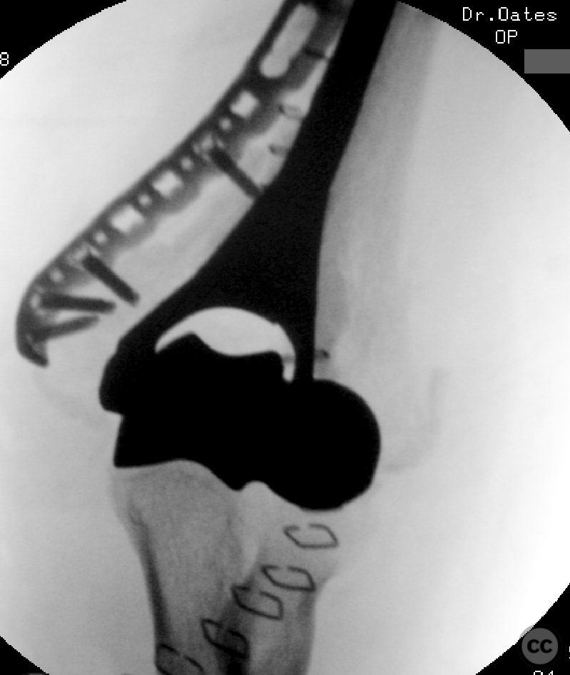
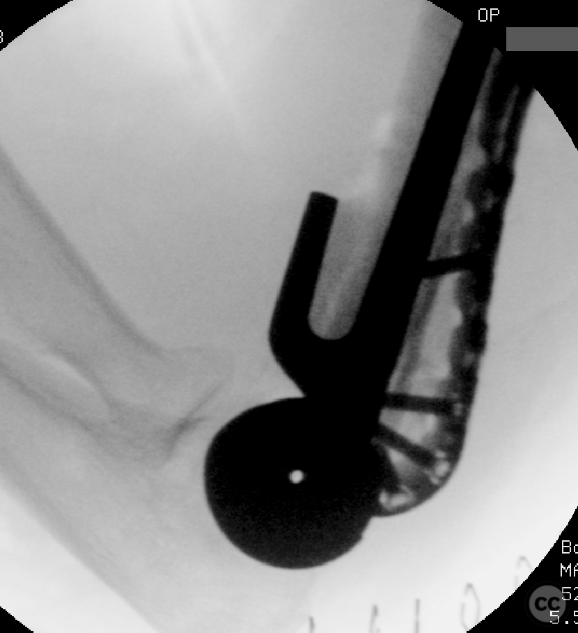
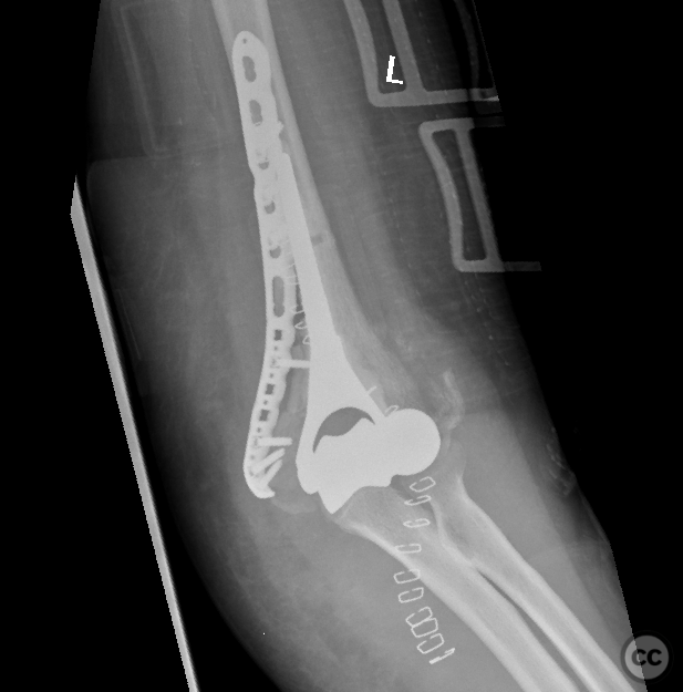
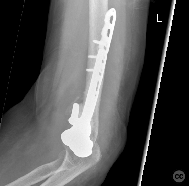
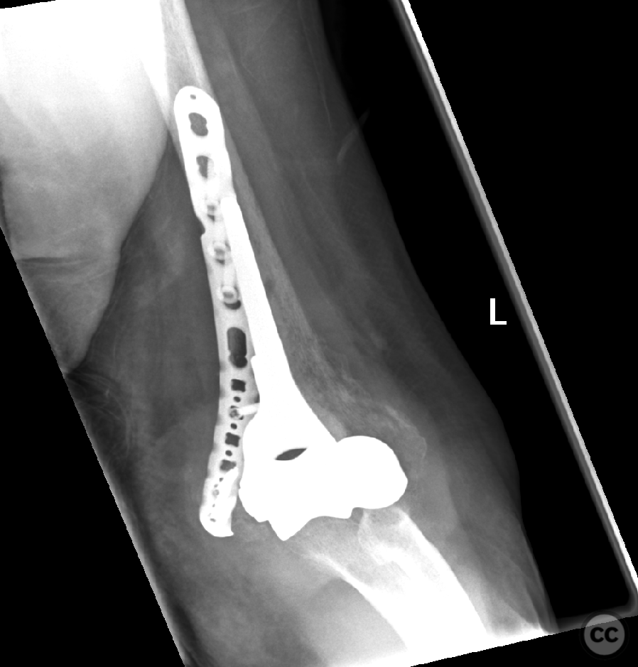
Clinical and radiological findings: A 70 year old lady sustained a low energy fall from standing height sustaining this isolated injury to the left elbow. A closed and neurovascular intact injury. The limb was placed in an above elbow cast to facilitate soft tissue consolidation, and allow the delivery of implants. The fracture constellation is best described as a highly comminuted Dubberly 4B articular distal humerus fracture. Whilst the goal of the majority of distal humerus articular injuries is anatomical restoration of joints surface with fragment specific osteosynthesis, this case presents the conundrum of a non-reconstructable injury in an elderly, but independently living moderately high functioning individual.
Preoperative Plan
Planning remarks:
Surgical Discussion
Operative remarks:In anticipation of a non-reconstructable situation , a Stryker Latitude arthroplasty system was organized. In addition, a full complement of osteosynthetic implants were available should the situation prove to be salvageable. The operation took place on day seven post injury. Patient was positioned supine with an arm board located across the chest upon which the flexed forearm could be placed. A midline posterior incision was made coursing radially around the olecranon. Full thickness flaps were developed mediately and laterally over the fascia of triceps. Identification and mobilisation of the ulnar withidentification of the first motor branch to FCU was identified. Proximal mobilization of the nerve to around 8 cm proximal to the joint. A transposition of the nerve was planned at this stage. Next we developed a medial paratricipital window and on the radial side, a radial paraolecranon window was established with extension to insertion of anconeus on the ulnar border. Through these two working windows a good view of the joint was established. It was identified early that it was a non-reconstructable injury. Removal of remarkable number of small fragments followed. A bony avulsion of the lateral collateral ligament complex allowed easy subluxation of the joint medially. This allowed unrestricted preparation of the distal ulnar humerus for implantation of the prosthesis. Unfortunately, during preparation of the box cut, the medial column broke away from the diaphysis. This whilst not hindering the final implantation of the prosthesis did compromise stability and required independent locking plate fixation. Further stabilization of both medial and lateral ligamental structures was established through suture fix ation through the cannulation of the spindle. Following ligamental reconstruction, a congruent range of motion without subluxation was demonstrable through a range of motion from 15 to 120°. The ulnar nerve, which originally was anticipated to be transposed consistently found its own way back into the ulnar groove posterior to the medial condyle. Therefore, a decision was made to leave it where it seemingly wanted to sit. There appear to be no impingement or contact of significance with prosthesis or osteosynthetic material. Postoperatively the ulnar nerve demonstrated a paresthesia without motor. Compromise. The paresthesia was reducing over the postoperative period. At six weeks I residual light paresthesia in ulnar nerve region was still identifiable. No motor deficits. Range of motion at time of review was zero 50,90° and stable.
Orthopaedic implants used: Tornier / Stryker Latitude
Author's Resources & References
Search for Related Literature

Dr Ed Oates
- Germany , Schleswig Holstein
- Area of Specialty - General Trauma
- Position - Specialist Consultant

Industry Sponsership
contact us for advertising opportunities



Article viewed 1032 times
09 Nov 2022
Add to Bookmarks
Full Citation
Cite this article:
Oates, E.J. (2022). Fracture hemiarthroplasty distal humerus. Journal of Orthopaedic Surgery and Traumatology. Case Report 19313014 Published Online Nov 09 2022.