


Schatzker III - arthroscopically assisted joint surface reduction
Score and Comment on this Case
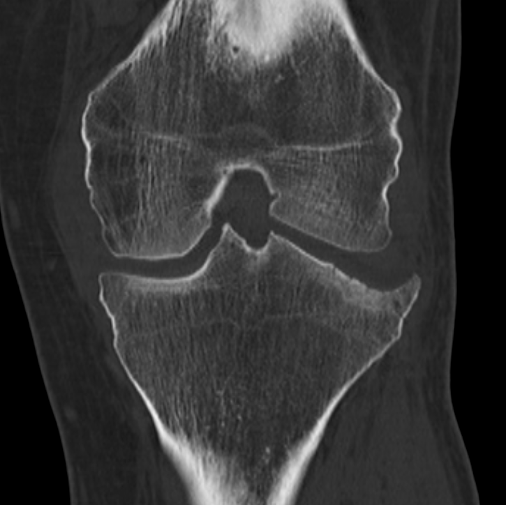
Clinical Details
Clinical and radiological findings: This case is a 48-year-old male pedestrian versus car who sustained a direct impact to the lateral aspect of the left knee. A combination soft tissue and bony injury he sustained: an incomplete rupture of the femoral aspect of the medial collateral ligament, a posterior horn tear of the medial meniscus and a Schatzker III lateral tibial plateau impression fracture. The pre-operative imaging included plain film XR, CT and MRI imaging whereby no further injury was identified, in particular both ACL & PCL presented radiologically intact. The lateral tibial plateau fracture was best described as an isolated impression with a maximal depression of 6 mm in the central aspect of the weight-bearing zone of the lateral plateau. After discussion the various management options with the patient we decided to manage the medial collateral ligament injury conservatively, and the medial meniscal injury and the lateral tibial plateau fracture would be managed simultaneously operatively. The operation took place eight days following the date of injury. Arthroscopic debridement of the medial meniscal injury was unremarkable. Then moving on to the lateral aspect of the joint the impression of the articular surface was clearly identifiable as demonstrated in the images above. An intra-articular depression of around 5 millimeters is clearly identifiable. Following arthroscopic identification of the injury we made a hockey stick skin incision over the lateral aspect of the proximal tibia. Inferior to Gerdy's tubercle we created a 1 cm cortical window through which we were able to decompress the fracture and reduce the articular surface to an anatomical level. The sub chondral bony defect was then filled with synthetic calcium phosphate bone void filler (ChronOs). Further impaction and compression of the synthetic material produced a satisfyingly dense support to the articular surface. Closure of the osteotomy window with the cortical flap (with periosteal hinge) was easily achieved - the placement of the lateral proximal locking plate simultaneously supported the closure of the window, and was used to create a raft screw construction under the chondral surface to further support the reduction against secondary compression / loss of reduction.
Preoperative Plan
Planning remarks:
Surgical Discussion
Operative remarks:This case was relatively straight forwards and the reduction of the articular surface is always satisfying to observe arthroscopically. Having seen secondary compression and loss of reduction of these fractures I’ve become increasingly rigorous with not only bringing a significant quantity of bone substitute into the void post reduction, but also with the compression of this synthetic material. The secondary raft screw construct could be achieved with individual screws plus an AP jail screw, however given the simplicity and stability of these proximal tibial LCP locking plates I generally prefer to bring the screws in through the plate.
Orthopaedic implants used: LCP Proximal Tibial Plate 3.5mm. ChronOS. Bone Graft Substitute
Author's Resources & References
Search for Related Literature

Dr Ed Oates
- Germany , Schleswig Holstein
- Area of Specialty - General Trauma
- Position - Specialist Consultant

Industry Sponsership
contact us for advertising opportunities
Article viewed 1755 times
24 Mar 2021
Add to Bookmarks
Full Citation
Cite this article:
Oates, E.J. (2021). Schatzker III - arthroscopically assisted joint surface reduction. Journal of Orthopaedic Surgery and Traumatology. Case Report 20802204 Published Online Mar 24 2021.