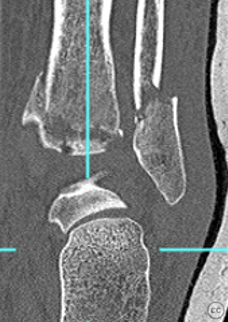
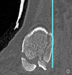
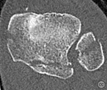
Mason and Molloy 2B Posterior Malleolar Fracture. Anterior collicular medial avulsion. Comminuted fibular
Score and Comment on this Case
Clinical Details
Clinical and radiological findings: Fracture Type - Lauge Hansen – SER 4 Mason and Molloy – 2B AO – 44B3.3 Soft -Tissue - Closed fracture Tscherne – C1 Specific Problems - Oblique comminuted fibular fracture. Type 2B PM fracture, anterior collicular fracture medially.
Preoperative Plan
Planning remarks: The 2B fracture pattern needs medial access to allow PM reduction first. We also need to ensure a comminuted fibular fracture can be accessed and reduced appropriately.
Surgical Discussion
Patient positioning: Recovery position.
Anatomical surgical approach: Lateral and medial posteromedial
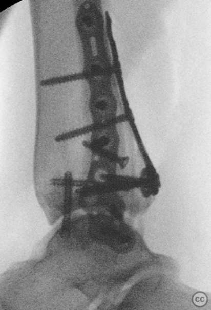
Operative remarks:My approaches were planned to allow confident reduction of the fibular through a direct lateral approach. Needing access to the PM and PL fragments the interval between tibialis posterior and FDL was most sensible. Therefore, a lateral and MPM is easiest in this case. The MPM can be curved anteriorly along the tib post course to allow access the anterior collicular fracture.
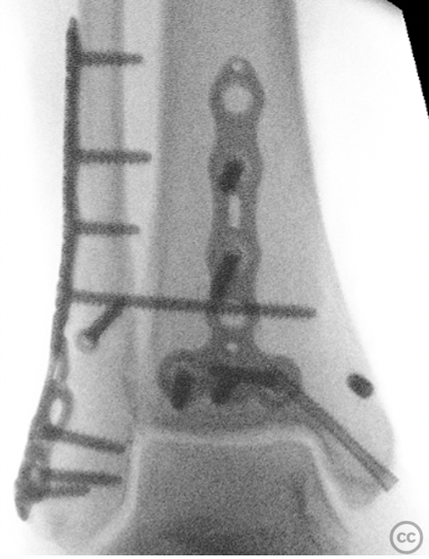
The MPM approach allowed me to reduce the PM fragment which was initially K wired in position. The PL fragment was then reduced and K wired in position before application of plate. The type 3 plate fitted best in this ankle with an additional headless PM screw to ensure reduction and rigid fixation of both fragments.
The fibular was redcued and I was able to lag before plate application. The anterior collisular fracture was reduced and fixed with a small headless screw. The syndesmosis was screened and as unstable was fixed with x1 screw. This is important as just fixing the PM fracture doesn't mean the anterior syndesmosis is not still unstable.
Postoperative protocol: 2 weeks NWB in a back slab. Conversion to Vacuum cast boot at 2 weeks to allow full weightbearing. Wean out of boot at 6 weeks.
Orthopaedic implants used: Volition (Orthosolutions), Accumed Headless 3.5mm screws
Search for Related Literature
Industry Sponsership
contact us for advertising opportunities



Article viewed 900 times
04 Feb 2023
Add to Bookmarks
Full Citation
Cite this article:
Mason, L. (2023). Mason and Molloy 2B Posterior Malleolar Fracture. Anterior collicular medial avulsion. Comminuted fibular. Journal of Orthopaedic Surgery and Traumatology. Case Report 20931103 Published Online Feb 04 2023.