
Bimalleolar Supination Adduction (SAd II) ankle injury
Score and Comment on this Case
Clinical Details
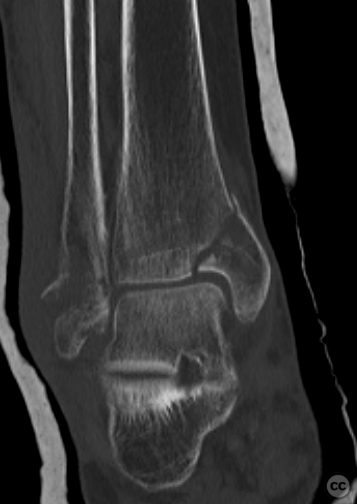
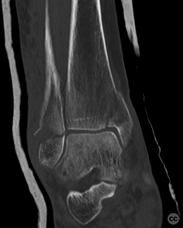
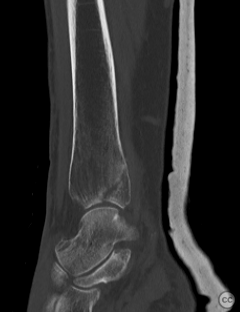
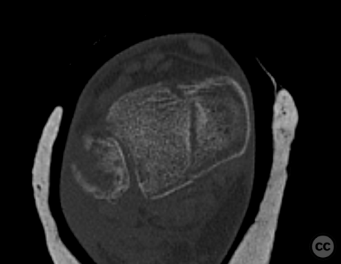
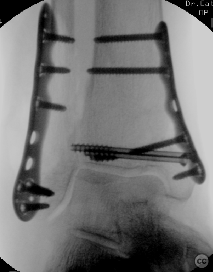
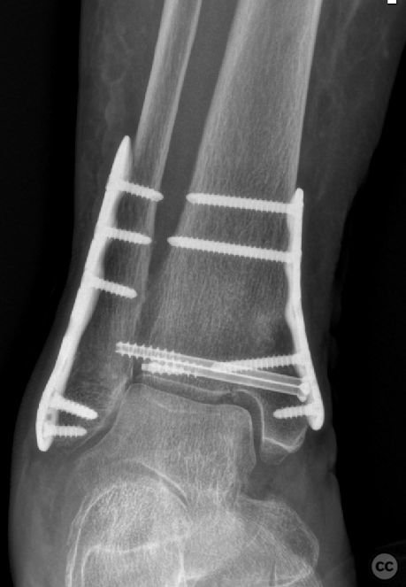
Clinical and radiological findings: 80yo F following a low energy supination-adduction (SAd II) injury with a Herscovici 4 vertical shear fracture of the medial malleolus and subsyndesmosal fracture of the distal fibula. The incidence of the supination adduction injury subtype is a relatively rare animal with an incidence of around 4 to 5% of bimalleolar fractures. Significant post-traumatic arthritis is quite high in this subgroup with the majority going on to have below average or poor functional outcomes. The inadequate reduction of marginal impaction is associated with an elevated risk for this post traumatic arthritis. The incidence of such a marginal plafond impression is around 60%.
Preoperative Plan
Planning remarks:
Surgical Discussion
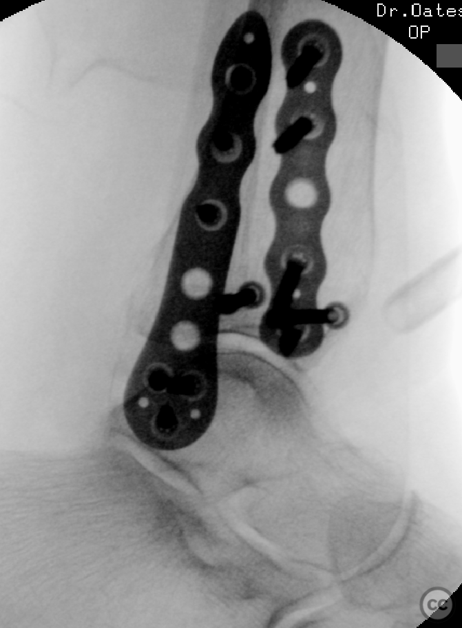
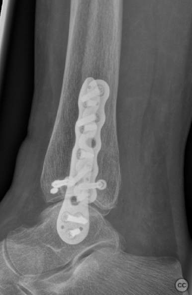
Operative remarks:Preoperative CT imaging suggested only minimal objective plafond impaction associated with this vertical medial malleolar fracture. A standard anteriomedial surgical approach was used and the fragments were primarily distracted allowing antegrade view through the fracture to the tibiotalar joint space. There was no objective indication of significant joint surface impaction. Accordingly no disimpaction was done. Following reduction of the fragment and temporary fixation a polyaxial fluoroscopic examination did not demonstrate an appreciable zone of impaction. Fixation of the medial malleolus was achieved primarily with the transverse lag screws with supplementary anti glide plate fixation overlapping the proximal fracture apex. Despite intraoperative clinical and radiological assessment, post-operative imaging does suggest some local impaction of the articular surface in this marginal medial region. I remain on the fence however given the corresponding shape of the medial talus shoulder and the apparent congruency of the joint space, I would consider the congruent curvature curvature demonstrated to be likely physiological.
Orthopaedic implants used: Stryker VariAx Distal Fibula
Author's Resources & References
Search for Related Literature

Dr Ed Oates
- Germany , Schleswig Holstein
- Area of Specialty - General Trauma
- Position - Specialist Consultant

Industry Sponsership
contact us for advertising opportunities
Article viewed 1185 times
27 Dec 2022
Add to Bookmarks
Full Citation
Cite this article:
Oates, E.J. (2022). Bimalleolar Supination Adduction (SAd II) ankle injury. Journal of Orthopaedic Surgery and Traumatology. Case Report 21617217 Published Online Dec 27 2022.