Mason II Radial Head Fracture
Score and Comment on this Case
Clinical Details
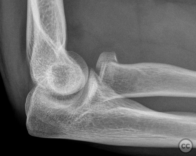
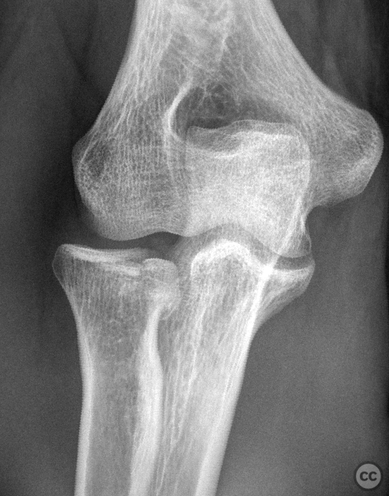
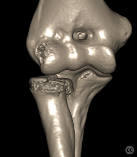
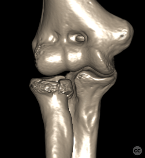
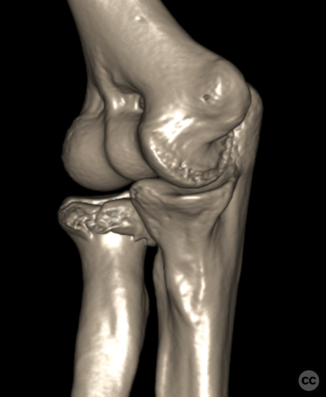
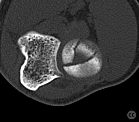
Clinical and radiological findings: A 24-year-old male presented after an e-scooter accident with a Mason II fracture of the radial head. Initial CT imaging revealed two discrete displaced fragments with approximately a 4mm intra-articular step. One fragment had extruded into the proximal radioulnar joint (PRUJ), causing an intermittent mechanical block to pronation.
Preoperative Plan
Planning remarks: The surgical plan involved a lateral approach with an extensor digitorum communis (EDC) split for minimally invasive Herbert Screw (HBS) screw fixation.
Surgical Discussion
Patient positioning: The patient was positioned supine with a tourniquet placed on the upper arm, followed by sterile preparation and draping of the operative field. A team timeout was conducted along with the administration of two grams of prophylactic antibiotics.
Anatomical surgical approach: A curved incision was made over the lateral epicondyle of the humerus extending distally over the radial head. The extensor digitorum communis muscle belly was identified, and its fibers were split to expose the joint capsule. A longitudinal incision of the capsule was made from the level of the capitulum, extending over the radial head into the proximal aspect of the radial neck. Anterior leaf of the capsule was lightly elevated from its insertion on the capitulum to enhance visualization of the anterior aspect of the joint.
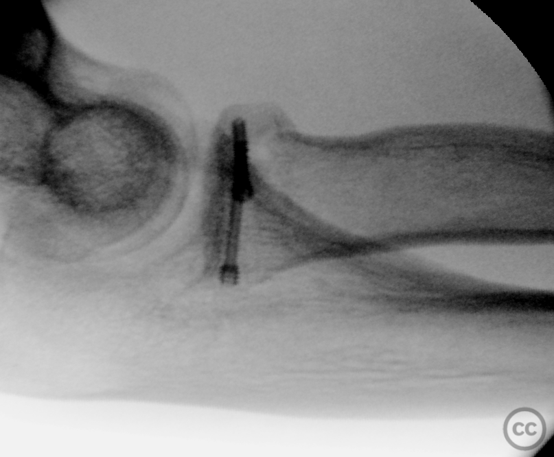
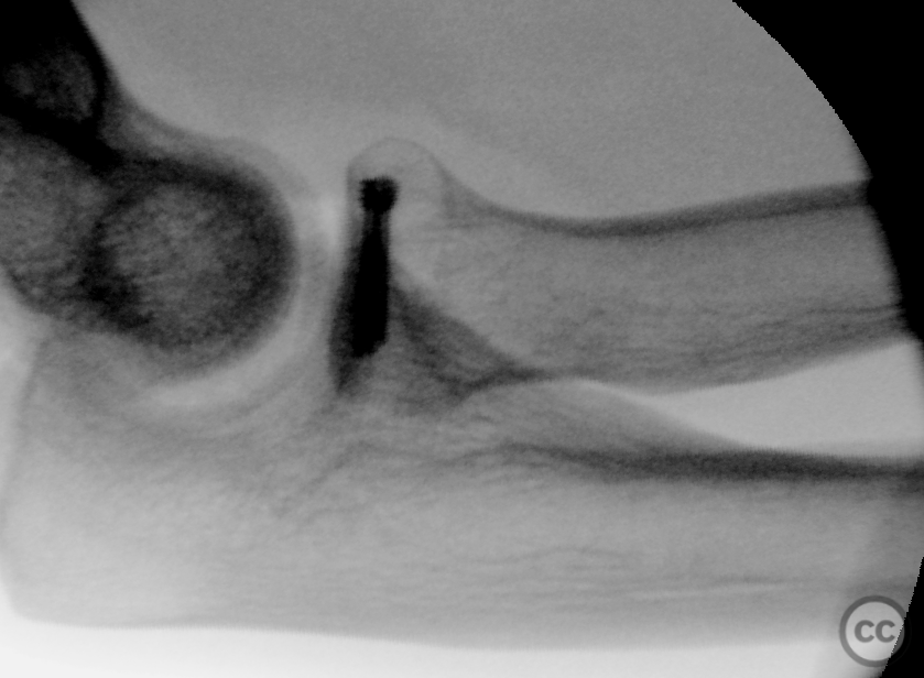
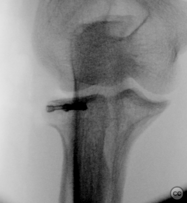
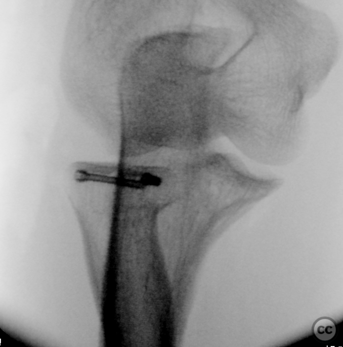
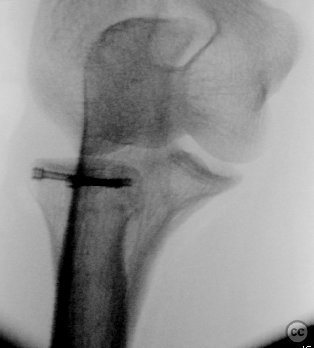
Operative remarks:After irrigation and hematoma removal, the two displaced fragments were located at the anterior and anteromedial aspects of the radial head. A 4mm osteotome was introduced into the fracture line, aiding in disimpaction and reduction of one fragment. A 2mm K-wire served as a bone tamp for the second fragment's reduction. Temporary fixation with a 0.8mm K-wire preceded the introduction of a single HBS Mini screw, uniting the fragments into a two-fragment construct. A second HBS Mini screw was inserted radially in subchondral bone, crossing the fracture plane at approximately 90 degrees, ensuring stepless articular reduction and stable osteosynthesis. Joint congruency was confirmed through 180 degrees of rotation, with fluoroscopy validating excellent reduction and restoration of articular axis.
Postoperative protocol: Immediate postoperative protocol allowed for free non-weight bearing range of motion in extension, flexion, supination, and pronation without the need for a splint.
Follow up: Not specified.
Orthopaedic implants used: Two HBS Mini screws.
Search for Related Literature

Dr Ed Oates
- Germany , Schleswig Holstein
- Area of Specialty - General Trauma
- Position - Specialist Consultant

Industry Sponsership
contact us for advertising opportunities
Article viewed 728 times
07 Mar 2024
Add to Bookmarks
Full Citation
Cite this article:
Oates, E.J. (2024). Mason II Radial Head Fracture. Journal of Orthopaedic Surgery and Traumatology. Case Report 23518827 Published Online Mar 07 2024.