Malunion Salter III distal radius fracture dislocation
Score and Comment on this Case
Clinical Details
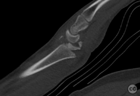
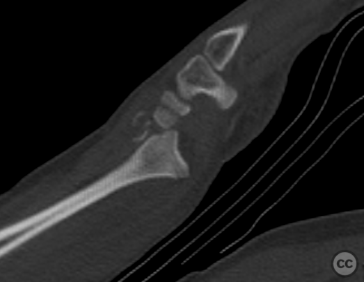
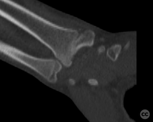
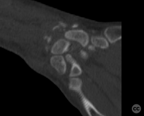
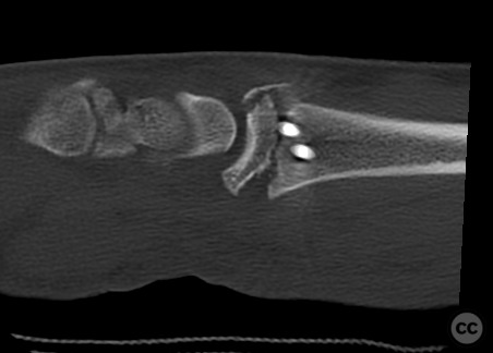
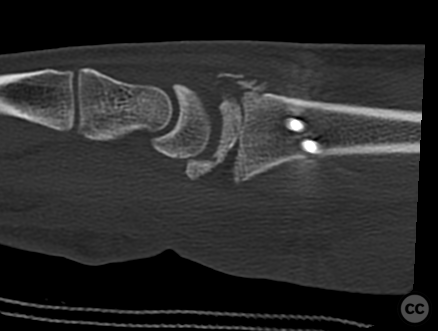
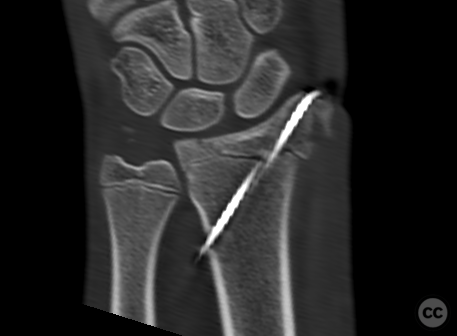
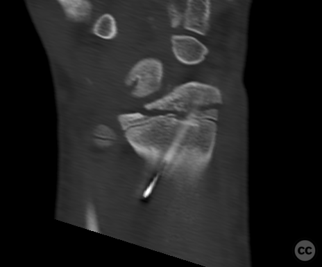
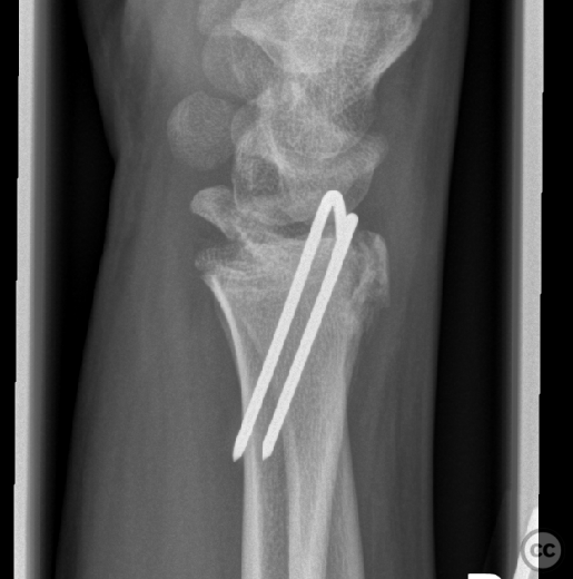
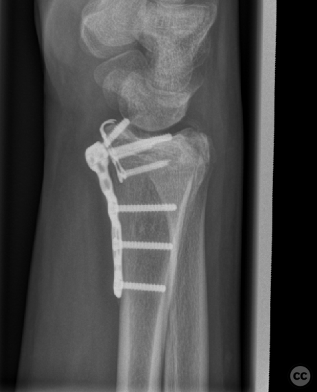
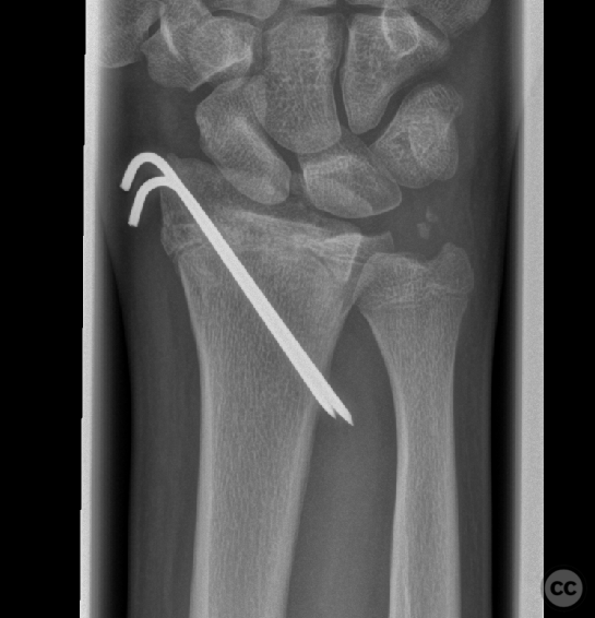
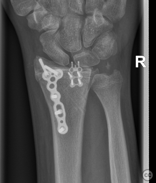
Clinical and radiological findings: This young teenager sustained a fall from over 3m directly onto the outstretched hand. This high energy injury resulted in a Salter III dorsal fracture dislocation of the distal radial epiphysis. Due to incomplete skeletal maturity the fracture demonstrated characters of a Tillaux distal tibia fracture with a transepiphyseal fracture line resulting in both retained and displaced articular surface elements. The index surgery was performaed by another surgeon and comprised of closed reduction and percutaneous k-wires over the radial styloid. The intra- and postoperative films, whilst demonstrating reduction of the dorsal carpal dislocation, also demonstrated an incomplete malreduction of the radial epiphyseal fracture. The significance of the malreduction was not appreciated and only at 5 weeks was the degree of malreduction appreciated by another surgical team. After discussion both within the surgical team and with patient and family a surgical correction at six weeks was planned with a correction osteotomy through the fracture and anatomic reduction. Given the small caliber of the volar rim fragment only few available plating systems would be capable of capturing and fixing the lunate fossa. Discussions with Medartis DE provided a solution with their 1.5 mm lunate hook plates in combination with their wrist system plates. The correction took place six weeks post index surgery. Modified Henry’s to the distal radius with extension across the wrist crease following flexor carpi radialis. Careful soft tissue release provided excellent view across the complete radial metaphysis. Using 0.5 mm oscillating blade on a small handpiece the original fracture plane was osteotomised under radiological guidance. Debridement of callus and soft tissue resulted in good mobilization of the fracture fragments without injury to the residual intact aspect of the lunate fossa. A small volar arthrotomy was performed through the radioscapholunate ligament providing view of reduction of the articular surface. This arthrotomy was also used for the insertion of the volar lunate hook plate and fixation with 1.5 mm screws. A dorsal radial plate was chosen to sit parallel to the ulna plate along the radial column providing locked screw fixation into the radial styloid. Given delicate the nature of the lunate plate hooks, suture fixation of the capsule directly to the plate was performed,, hereby providing both closure of the arthrotomy and reinforcement of capsular attachment stress-relieving the plate hooks.
Preoperative Plan
Planning remarks:
Surgical Discussion
Operative remarks:There is a paucity of literature to guide treatment and timing of malreduction/malunion in this injury constellation. Given the incomplete involvement of the articular surface an en bloc corrective osteotomy at a later time was considered insufficient to reduce intra-articular joint surface step off. A fragment specific osteotomy was required. A correction still winthin the early phase of bone healing was considered to provide the best opportunity to achieve fragment specific mobilization and reduction. Given the patient’s young age any remaining remodelling capacity could also be used to our advantage.
Orthopaedic implants used: Medartis Aptus Wrist System
Author's Resources & References
Search for Related Literature

Dr Ed Oates
- Germany , Schleswig Holstein
- Area of Specialty - General Trauma
- Position - Specialist Consultant

Industry Sponsership
contact us for advertising opportunities




Article viewed 1237 times
09 Nov 2022
Add to Bookmarks
Full Citation
Cite this article:
Oates, E.J. (2022). Malunion Salter III distal radius fracture dislocation. Journal of Orthopaedic Surgery and Traumatology. Case Report 25691878 Published Online Nov 09 2022.