


Distal Radius Fracture with Lunate Facet Involvement and Radiocarpal Dislocation
Score and Comment on this Case
Clinical Details
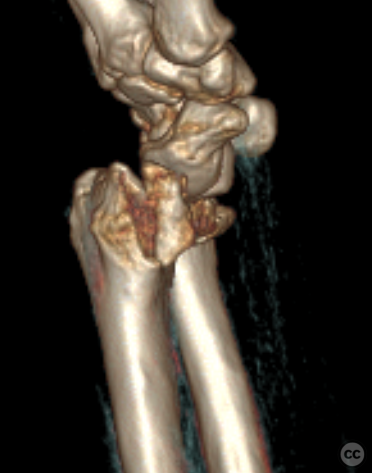
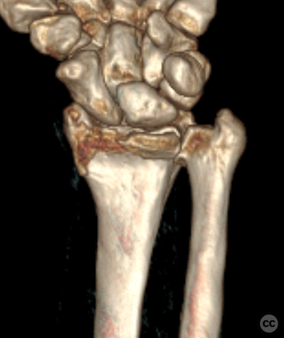
Clinical and radiological findings: A 71-year-old male presented following a trip and fall from standing height, resulting in a palmar radiocarpal dislocation and an associated distal radius fracture. Initial radiographs and CT imaging confirmed the fracture dislocation, specifically demonstrating involvement of the lunate facet and a coronal split of the radial styloid. Marked radiocarpal arthritis, preexisting radial shortening, and increased volar angulation were noted, suggesting a previous distal radius fracture, although the patient was unsure of any prior injury.
Preoperative Plan
Planning remarks: plan
Surgical Discussion
Patient positioning: The patient was positioned supine with the left arm placed on an auxiliary arm table.
Anatomical surgical approach: A longitudinal incision was made over the flexor carpi radialis tendon, extending across the wrist crease in a zigzag fashion, approximately nine centimeters in length. Subcutaneous dissection identified the flexor carpi radialis tendon, which was retracted ulnarly after incising its sheath. The antebrachial fascia was incised to reveal the flexor pollicis longus, which was also retracted ulnarly. The pronator quadratus was incised along its radial border and along the watershed line, with ulnar retraction of the muscle belly. This exposed a volar rim fracture extending from the radial styloid to the ulnar lunate facet, with subluxation of the carpus in the volar direction.
Operative remarks:Debridement and irrigation of the fracture gap and radiocarpal joint were performed. Reduction of the lunate facet and volar rim fragment was achieved and provisionally fixed with a 1.0 millimeter K-wire. Definitive fixation was secured with an HB’s mini headless compression screw. Due to insufficient mechanical stability, a 2.0 millimeter striker mini-fragment plate was cut and bent into a volar rim hook plate, secured with two 2.4 millimeter bicortical screws. Attention was then directed to the radial styloid fracture, which was reduced using a Hinterman K-wire distractor after overcoming brachioradialis tension. A 2.0 millimeter striker mini-fragment plate was placed and secured with a bicortical 2.4 millimeter screw, three distal subchondral screws, and two further bicortical proximal screws. Multiplanar fluoroscopy confirmed anatomical reduction and satisfactory hardware placement without residual radiocarpal instability. Irrigation with normal saline solution was performed, followed by adaptation of the pronator quadratus muscle over the plates for protection of the flexor pollicis longus tendon. Closure of subcutaneous tissue and skin was achieved with running sutures, followed by application of a sterile dressing and elastic bandage.
Postoperative protocol: Free ROM exercises for 6W pripr to commencement of weight-bearing
Follow up: Not specified
PubMed® Literature Review
Generated by The Literature - Realtime PubMed® AnalysisHook plate fixation of volar lunate rim fragments in distal radius fractures offers stable anatomical fixation with favorable functional outcomes. Despite a high rate of hardware-related complications, advancements in plate design have mitigated these issues. Overall, hook plate fixation is a viable option for managing volar marginal rim fractures, particularly when fragment-specific fixation is required.
Evidence and Analysis:
The management of volar lunate rim fragments in distal radius fractures presents significant challenges due to the intricate anatomy and the need for precise fragment stabilization. Hook plate fixation has emerged as a specialized technique addressing these challenges. O'Shaughnessy et al. (2015) conducted a Therapeutic IV study involving 26 wrists, demonstrating that volar fragment-specific hook plates provide stable fixation without loss of the critical volar ulnar corner [4]. This study reported no instances of carpal subluxation, underscoring the plate's efficacy in maintaining anatomical alignment. However, hardware irritation was noted in five patients, primarily associated with second-generation plate designs featuring prominent bends, leading to the necessity of hardware removal in four cases.
Further supporting these findings, Gavaskar et al. (2021) evaluated 18 patients with volar marginal fragments managed using an anatomical volar hook plate alongside conventional volar locking plates [9]. All fractures achieved union with maintained reduction of the volar marginal fragment and stable radiocarpal and distal radioulnar joints. Functional outcomes were favorable, with a mean Mayo wrist score of 75 and PRWE score of 15.2, indicating significant wrist function recovery. However, the study acknowledged potential complications related to implant prominence, which necessitates careful plate selection and placement.
Bakker and Shin (2014) presented an early exploration of fragment-specific volar hook plates in six cases, reporting no surgical complications and successful stabilization of the volar lunate rim fragments [11]. This preliminary evidence suggested that such plates could effectively address the limitations of standard volar locking plates in capturing small, critical fragments. Similarly, Kachooei et al. (2016) assessed ten patients treated with the DePuy-Synthes Volar Rim Plate, achieving fracture healing in all cases with minimal motion deficits [13]. Nonetheless, one patient required secondary surgery for ulnar impingement, highlighting the risk of hardware-related issues.
Imatani and Kondo (2021) introduced the K-I classification and the PD technique for managing volar lunate facet rim fractures, focusing on the use of anatomical low-profile volar locking plates [7]. In their retrospective review of 35 patients, all fractures healed with good functional outcomes, as evidenced by an average Mayo wrist performance score of 81.7 and quick-DASH score of 9.5. The study emphasized that standard volar plates are insufficient for these fragments, thereby necessitating specialized fixation methods like the PD technique to prevent complications such as radiocarpal subluxation and malunion.
Lari et al. (2023) conducted a systematic review of operative treatments for volar rim fractures, encompassing 26 studies with 617 wrists [2]. The review highlighted that standalone hook plates were among the most commonly utilized implants (13%) and reported an overall complication rate of 14%, with a significant portion related to flexor tendon problems. The implant removal rate was 22%, indicating a substantial need for secondary interventions. Despite these complications, functional outcomes across various treatment modalities, including hook plates, were generally favorable.
Stein et al. (2024) explored the necessity of repairing acute scapholunate injuries in the context of distal radius fractures, indirectly relevant to the discussion of volar rim fixation [3]. While not directly addressing hook plates, the study's findings on ligamentous stability contribute to the broader understanding of carpal stability post-fixation.
Tordjman et al. (2016) proposed the extensile volar-ulnar approach for fixing volar lunate facet fragments, offering an alternative to traditional volar approaches that may limit fragment exposure [6]. This technique facilitates optimal reduction and stabilization, complementing hook plate fixation by ensuring comprehensive fragment management.
Lastly, Wiesler et al. (2006) described an arthroscopic technique for managing volar lunate facet fractures [8]. Although not directly related to hook plates, the minimally invasive approach emphasizes the importance of accurate fragment visualization and reduction, principles that underpin effective hook plate fixation.
Collectively, the evidence underscores the efficacy of hook plate fixation in providing stable, anatomically accurate fixation of volar lunate rim fragments. The high union rates and favorable functional outcomes reported across studies attest to the technique's effectiveness. However, the consistent occurrence of hardware-related complications, particularly tendon irritation, warrants attention. Advances in plate design, such as lower-profile hooks and modified bends, have mitigated some of these issues, as evidenced by reductions in hardware irritation post-design revisions [4]. Additionally, the integration of fragment-specific approaches like the PD technique [7] and extensile volar-ulnar exposures [6] enhances the stability and functional restoration achieved with hook plates.
The quality of evidence, primarily comprising Therapeutic IV studies and systematic reviews, limits the ability to perform high-level meta-analyses. The retrospective nature of most studies introduces potential biases, and the heterogeneity in patient populations and surgical techniques complicates direct comparisons. Nevertheless, the convergence of findings across diverse studies reinforces the clinical utility of hook plate fixation for volar lunate rim fragments.
Discussion:
Hook plate fixation serves as a robust method for addressing volar lunate rim fragments in distal radius fractures, offering precise anatomical stabilization essential for optimal functional recovery. The reviewed literature consistently demonstrates high union rates and satisfactory wrist function post-fixation. The specialized design of hook plates caters to the unique anatomical challenges posed by small, critical fragments, ensuring their securement and preventing complications such as carpal subluxation and malalignment.
Despite these advantages, hardware-related complications, particularly tendon irritation, remain a concern. However, advancements in plate design, including lower-profile hooks and elimination of prominent bends, have effectively reduced such issues, as evidenced by the decreased need for hardware removal in more recent studies [4, 9]. The integration of fragment-specific techniques and comprehensive surgical approaches further enhances the stability achieved by hook plates, addressing both bone and soft tissue considerations.
The current evidence, while supportive of hook plate fixation, is predominantly derived from lower-level studies, highlighting the need for higher-quality research to substantiate these findings. Prospective, randomized controlled trials would provide more definitive evidence regarding the efficacy and safety of hook plates compared to alternative fixation methods.
In conclusion, hook plate fixation of volar lunate rim fragments is a viable and effective strategy in the surgical management of distal radius fractures involving these critical fragments. The technique's ability to provide stable, anatomical fixation with favorable functional outcomes positions it as a preferred option, particularly in complex fracture patterns where standard volar plates fall short. Ongoing refinements in plate design and surgical techniques are anticipated to further enhance the outcomes and reduce complications associated with this fixation method.
Search for Related Literature

Dr Ed Oates
- Germany , Schleswig Holstein
- Area of Specialty - General Trauma
- Position - Specialist Consultant

Industry Sponsership
contact us for advertising opportunities
User Discussion (1)
Guest User
Beautifully solved with clever solution
thanks! it was a good bailout, and all behaved nicely