.png)
.png)
.png)
.png)
.png)
.png)
.png)
.png)
.png)
.png)
.png)
.png)
.png)
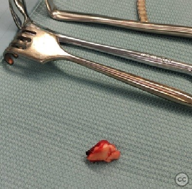
Trimalleolar ankle fracture with intermediate, posterior malleolus fracture fragment displaced to medial malleolus fracture gap.
Score and Comment on this Case
Clinical Details
Clinical and radiological findings: Clinical and radiological findings: Closed, Lauge-Hansen – SER 4 fracture (lateral malleolus - Weber A; posterior malleolus: Mason and Molloy – 2A with intermediate fragment displaced to medial malleolus fracture gap; transverse medial malleolus fracture). Special consideration: synostosis of the anterior part of the syndesmosis was probably the cause of an atypical lateral malleolar fracture morphology.
Preoperative Plan
Planning remarks: 1. Lateral position - removal of the posterior malleolus intermediate fragment from the medial malleolus fracture gap using a medial approach - direct lateral approach to the lateral malleolus - reduction of the posterior malleolus intermediate fragment through the lateral malleolus fracture gap and temporary fixation of the intermediate fragment using the lost k-wire technique - lateral malleolus anatomical reduction - percutaneous fixation of posterior malleolus in PA direction 2. Supine position - medial malleolus ORIF with two bicortical screws
Surgical Discussion
Patient positioning: Lateral, supine
Anatomical surgical approach: Direct lateral, medial, percutaneus for PM fixation
Operative remarks:I always perform the external rotation stress test under direct visual control of the anterior syndesmosis after fixation of the posterior malleolus. In most cases, after fixation of the posterior malleolus, the syndesmosis is still unstable in the direction of external rotation.
In this case, the anterior syndesmosis was undamaged due to its synostosis and the atypical morphology of the lateral malleolus fracture.
The atypical morphology of the lateral malleolus fracture meant that fixation of this fracture with a posterolateral plate alone did not provide complete stability, so I decided to place another plate on the lateral side.
Postoperative protocol: Weight-bearing as tolerated in a walker and ROM exercises after wound healing.
Follow up: AOFAS 87, OMAS 90 six months after surgery. 1 year after surgery, there was no need for implant removal (no signs of soft tissue irritation on the lateral side)
Orthopaedic implants used: Double Medical (lateral and postero-lateral plates
Author's Resources & References
Search for Related Literature


Industry Sponsership
contact us for advertising opportunities
User Discussion (1)
Guest User
great case, and nice fix. recently double-plated a fibula myself, no no words of criticism here (i probably should post that case!)