Corrective Osteotomy and Volar Plating for Malunited Distal Radius Fracture
Score and Comment on this Case
Clinical Details
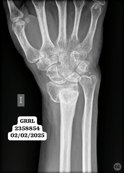
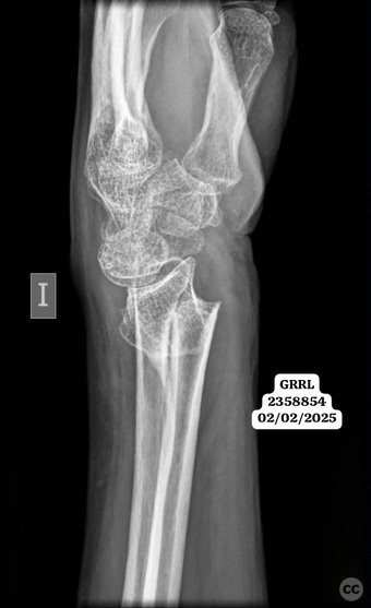
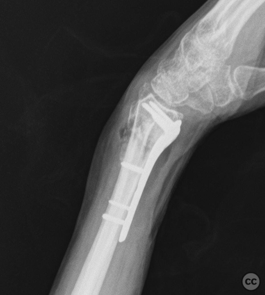
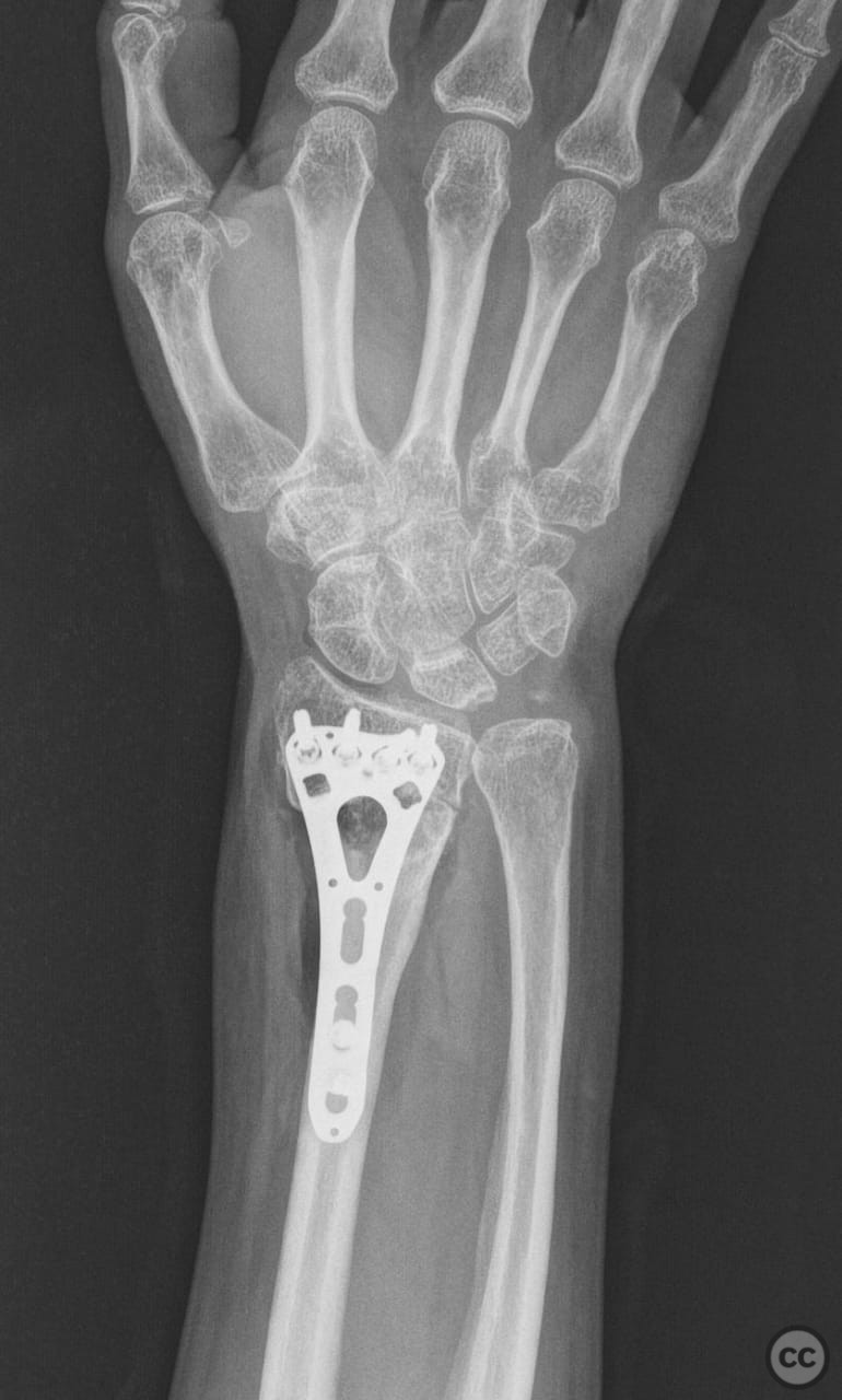
Clinical and radiological findings: A 59-year-old female presented with persistent pain and deformity of the left wrist, five months post-injury. Radiographic evaluation revealed a malunited distal radius fracture characterized by severe shortening and dorsal angulation. The fracture had consolidated in this malaligned position, necessitating surgical intervention.
Preoperative Plan
Planning remarks: The preoperative plan involved a modified Henry approach to the distal radius. The surgical strategy included performing a corrective osteotomy at the level of the deformity, parallel to the articular surface, followed by fixation with a volar locking plate. An autologous iliac crest bone graft was planned to address the dorsal defect.
Surgical Discussion
Patient positioning: The patient was positioned supine on the operating table with the left arm extended on a hand table to facilitate access to the volar aspect of the wrist.
Anatomical surgical approach: A modified Henry approach was utilized, involving an incision along the flexor carpi radialis tendon. Dissection proceeded between the flexor carpi radialis and the radial artery, with careful retraction of the flexor tendons and protection of the median nerve. The pronator quadratus muscle was elevated subperiosteally to expose the distal radius.
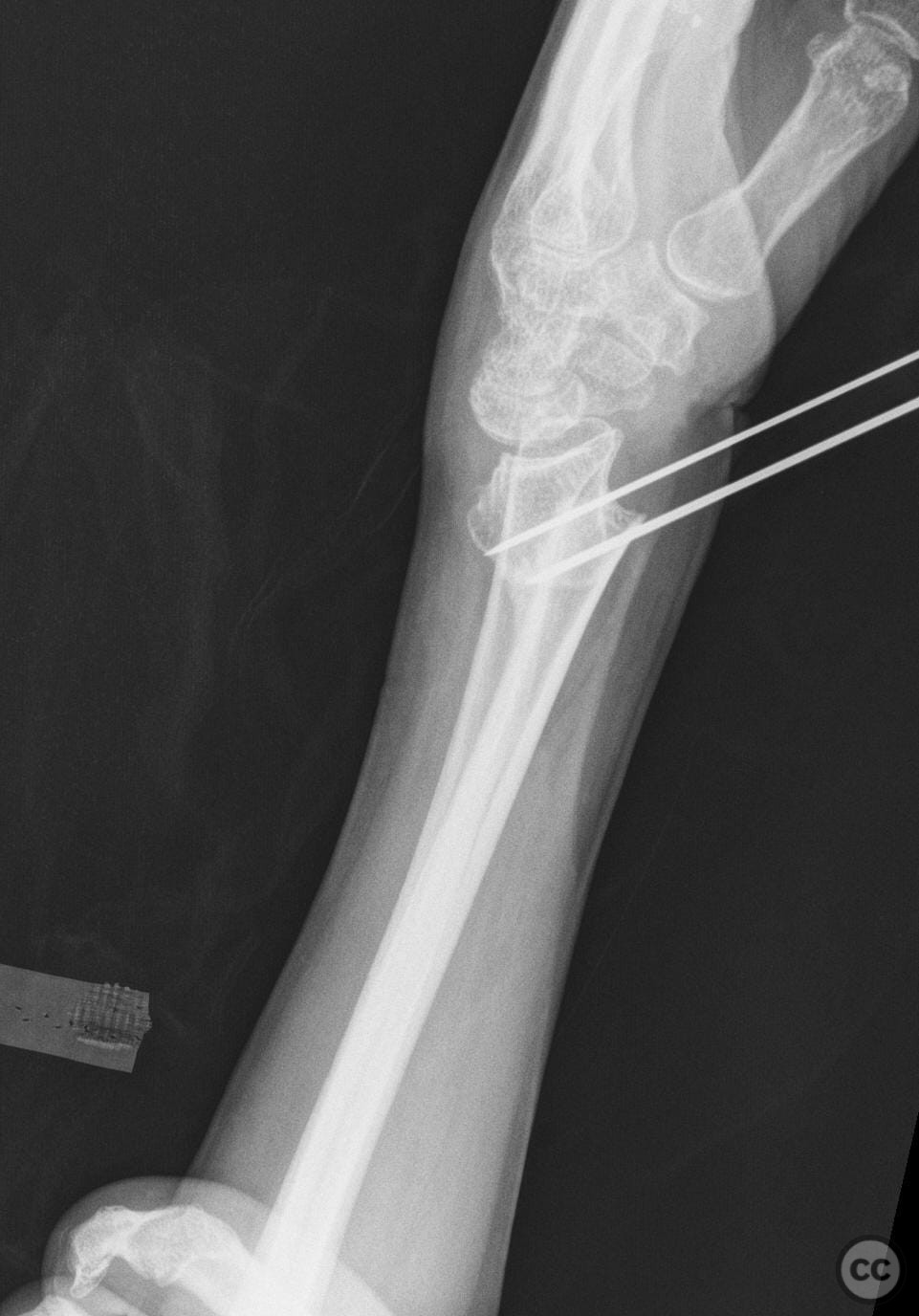
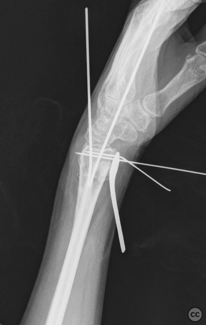
Operative remarks:Intraoperatively, a transverse osteotomy was performed at the site of deformity using an osteotome. Provisional fixation was achieved with Kirschner wires, allowing for realignment of the distal fragment. A volar locking plate was then applied distally to secure the reduction. The dorsal defect was filled with an autologous iliac crest bone graft to support bone healing and restore radial length.
Postoperative protocol: Postoperatively, the patient was immobilized in a volar splint for two weeks, followed by initiation of range of motion exercises. Weight-bearing activities were restricted for six weeks to allow for graft incorporation and fracture healing.
Follow up: Not specified.
Orthopaedic implants used: Volar locking plate, Kirschner wires, autologous iliac crest bone graft.
Search for Related Literature


Industry Sponsership
contact us for advertising opportunities
Article viewed 166 times
04 Feb 2025
Add to Bookmarks
Full Citation
Cite this article:
LUIS LEONCIO TEMOCHE DIAZ. (2025). Corrective Osteotomy and Volar Plating for Malunited Distal Radius Fracture. Journal of Orthopaedic Surgery and Traumatology. Case Report 30581946 Published Online Feb 04 2025.