
A case of Combined mechanism Pelvic ring injury-APC+LC
Score and Comment on this Case
Clinical Details
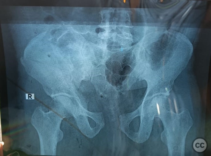
Clinical and radiological findings: 44/Male BMI-30 closed injury No Morel lavallee Neurovascular intact Xray-Pubic symphysis diastasis >2.5cm (APC) -Rt iliac wing fracture-exiting anterior to SI joint but involving.(LC) CT scan-Confirms xray diagnosis-no involvement of SI joint.
Preoperative Plan
Planning remarks: Planned to treat the injury by operative fixation as its an unstable pattern. Symphysis-open reduction and plate fixation Iliac wing-open reduction and plate fixation
Surgical Discussion
Patient positioning: Supine with a bump under right hip
Anatomical surgical approach: 1)Pfannensteil approach to reduce and fix symphysis,2)Lateral window approach to reduce and fix iliac wing.
Operative remarks:The symphysis reduction was achieved with traction,adduction,internal rotation of both lower limbs and by direct manual pressure of both greater trochanters.
Reduction was secured with a pointed reduction clamp and provisionally fixed with a kwire across symphysis.
6 holed pubic symphysis specific plate was placed and secured with k-wires.
Iliac wing fracture was exposed through lateral window approach.
Reduction was achieved indirectly during reduction of symphysis with some residual displacement which was reduced with a ball spike pusher and provisionally fixed with k-wires.
Definitive fixation of iliac wing fracture was achieved by interfragementary screws and a 6 holed curved pelvic recon plate.
Symphysis plate is fixed with 3 screws on left side and 2 screws on right as the last plate hole on right was off anteriorly.
Fixation strength was checked by stressing under continuous flouroscopy and found to be stable.
Wounds were closed with a drain after thorough saline lavage.
Postoperative protocol: Post op was uneventful. Patient was made to sit immediate post op day 1 passive hip ROM and active knee and ankle ROM started. Weight bearing restriction for 6 weeks Ambulated with walker after 6 weeks-weaning of until 3 months post op
Follow up: At 6 months follow up Wound healthy Full hip ROM Walking without any difficulty No restriction on daily activities No sexual dysfunction reported Patient was lost to follow up after that
Orthopaedic implants used: 6 holed symphysis plate 6 holed curved recon plate Interfragmentary cortical screws.
Search for Related Literature
Vishwanath Mahadevuni
- India , Hyderabad
- Area of Specialty - General Trauma
- Position - Specialist Consultant

Industry Sponsership
contact us for advertising opportunities
Article viewed 630 times
19 Jul 2024
Add to Bookmarks
Full Citation
Cite this article:
Vishwanath Mahadevuni. (2024). A case of Combined mechanism Pelvic ring injury-APC+LC. Journal of Orthopaedic Surgery and Traumatology. Case Report 43248336 Published Online Jul 19 2024.