Pediatric Femoral Shaft Fracture Managed with Elastic Stable Intramedullary Nailing (ESIN)
Score and Comment on this Case
Clinical Details
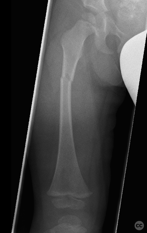
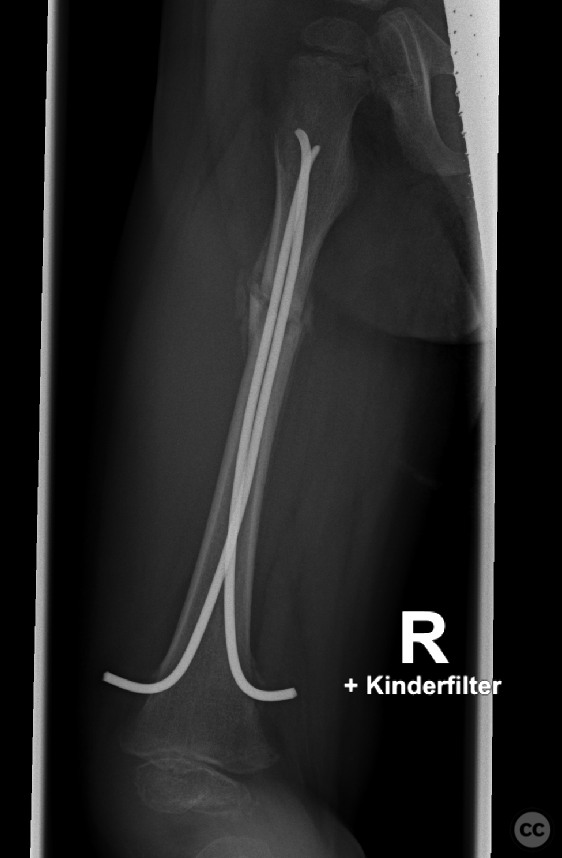
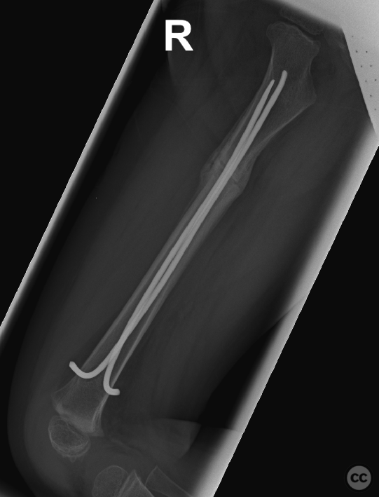
Clinical and radiological findings: A 4-year-old boy presented with pain in the right thigh after falling onto an edge from a slide at kindergarten. Clinical examination revealed localized tenderness and swelling over the right femoral shaft, with no evidence of open wounds or neurovascular compromise. Radiographs of the right femur demonstrated a ptoximal 1/3 shaft fracture with slight displacement but no comminution, consistent with a simple transverse pattern, which can be classified as a 32-A1 AO/OTA
Preoperative Plan
Planning remarks: The preoperative plan included closed reduction and internal fixation using elastic stable intramedullary nailing (ESIN) to provide stable fixation while allowing for early mobilization.
Surgical Discussion
Patient positioning: The patient was positioned supine on a radiolucent operating table to facilitate intraoperative imaging with a C-arm fluoroscope.
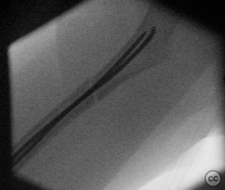
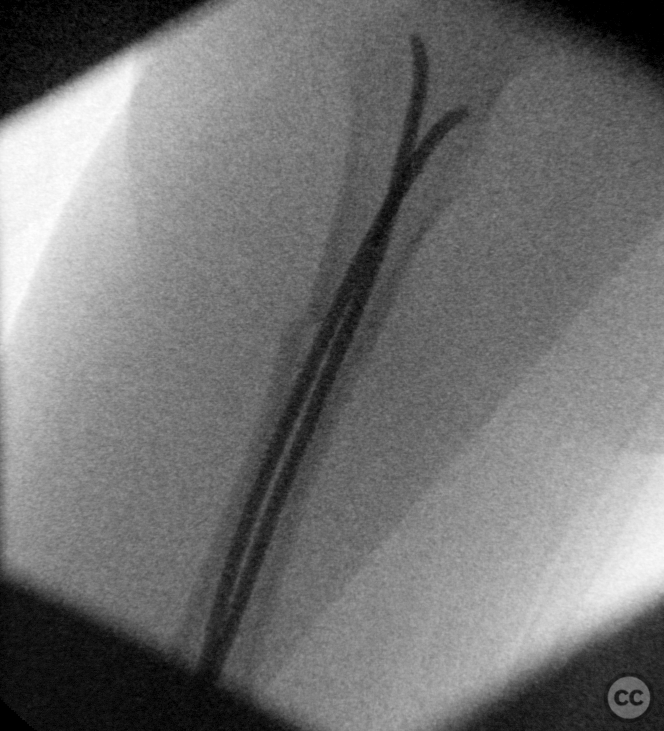
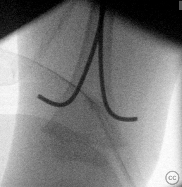
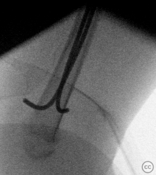
Anatomical surgical approach: A medial distal thigh incision was made to access the femoral medullary canal. The canal was opened proximal to the growth plate using an awl, ensuring no damage to the physis. A pre-bent titanium elastic nail was then inserted and advanced across the fracture site under fluoroscopic guidance until it reached the proximal fragment. A similar approach was used on the lateral side to insert a second nail, achieving bicortical purchase and ensuring proper alignment and rotation.
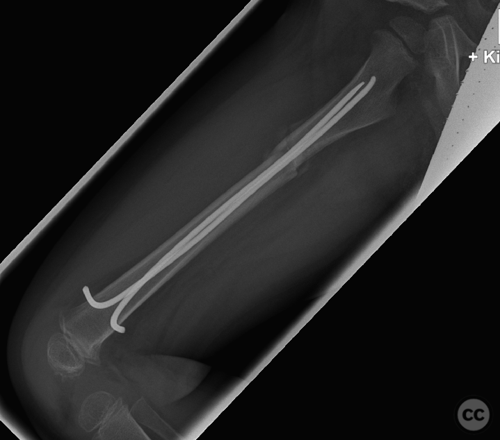
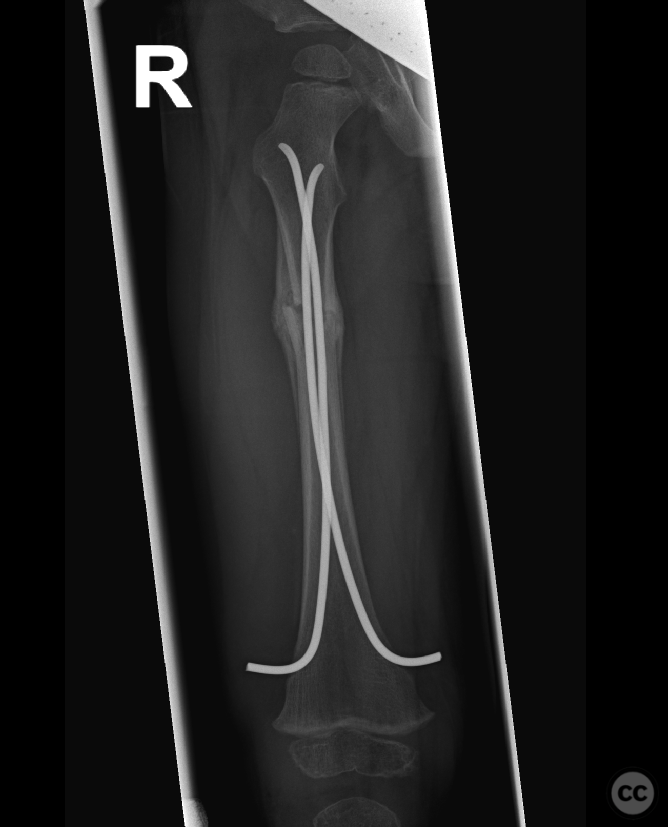
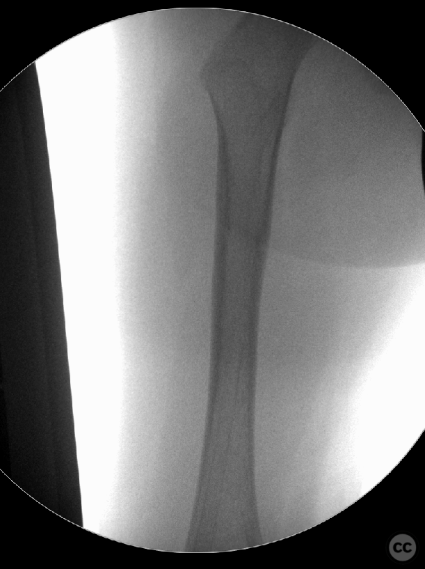
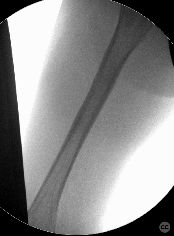
Operative remarks:Intraoperatively, the fracture was slightly distracted initially, but was successfully compressed after nail insertion to minimize the gap, achieving good axial alignment. The nails were bent and cut below skin level to prevent soft tissue irritation. The rotational alignment was confirmed with fluoroscopy, as wel as clinically comparing both legs to ensure symmetry.
Postoperative protocol: Postoperative management involved functional, pain-oriented rehabilitation without the need for immobilization. Weight-bearing as tolerated was encouraged based on the child's comfort level.
Follow up: Clinical follow-up at 6 months postoperatively showed satisfactory healing of the fracture with no signs of malalignment or leg length discrepancy. The ESINs were removed at this time, and the patient was able to resume normal activities without restrictions.
Orthopaedic implants used: Two titanium elastic stable intramedullary nails (ESIN)
Search for Related Literature

Dr Ed Oates
- Germany , Schleswig Holstein
- Area of Specialty - General Trauma
- Position - Specialist Consultant

Industry Sponsership
contact us for advertising opportunities
Article viewed 389 times
16 Feb 2024
Add to Bookmarks
Full Citation
Cite this article:
Oates, E.J. (2024). Pediatric Femoral Shaft Fracture Managed with Elastic Stable Intramedullary Nailing (ESIN). Journal of Orthopaedic Surgery and Traumatology. Case Report 46601443 Published Online Feb 16 2024.