Nonunion MT5 Zone 2 plus acute subcapital MT2/3/4 fractures
Score and Comment on this Case
Clinical Details
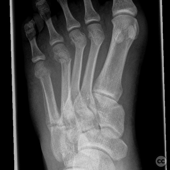
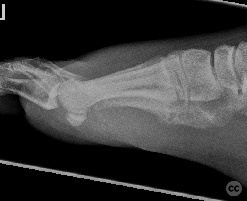
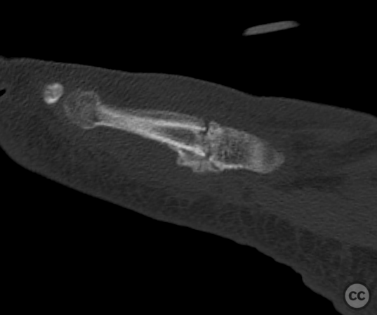
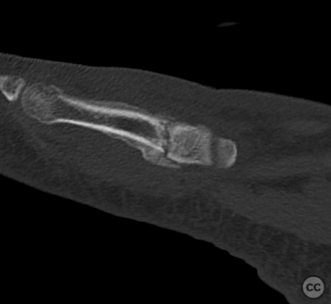
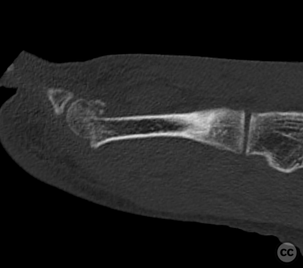
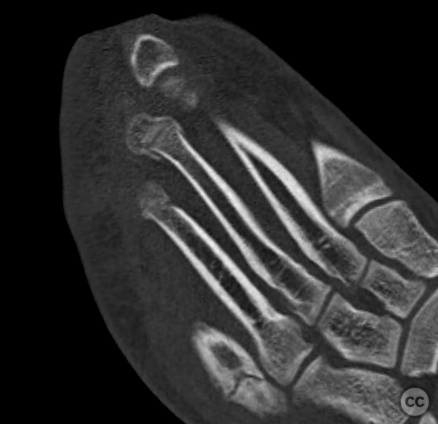
Clinical and radiological findings: This is a 30-year-old gentleman who presented to emergency following a motor vehicle accident. Plain film imaging demonstrated acute fractures of the metatarsal heads of the second, third, and fourth rays. Additionally a zone 2 fracture of the base of the fifth metatarsal is identifiable with evidence of extensive callus growth along the lateral metatarsal border. Further exploration of the history elucidated a sporting injury approximately three months prior with persisting pain on weight-bearing up to the time of current presentation. Plain film imaging was supplemented with CT which demonstrated marked dorsal displacement of the fourth metatarsal head along with delayed union of the Jones fracture and clear residual fracture line. Fractures of the 2nd and 3rd rays were minimally impacted and deviated minimally from physiological axis. Following discussion with the patient operative intervention was decided to reduce dorsal translation of the fourth ray. Percutaneous screw fixation was offered for the fifth ray in the same setting. The operation took place one week post injury. The patient was positioned supine on a radiotranslucent operating table. An above knee tourniquet was applied but not inflated. A percutaneous 4.5 mm cannulated partially threaded screw was brought in antegrade through the base of the fifth metatarsal crossing the fracture line and engaging the medial cortex of the diaphysis. This screw provided considerable compression at the fracture site which was witnessed on intra-operative fluoroscopy as closing of the residual fracture gap. The operation then continued with a small 5 mm incision over the proximal diaphysis of the fourth metatarsal. Opening of the metatarsal diaphysis was achieved using a 1.8 mm K wire. Through this 1.8 mm hole a 1,6mm K wire was then introduced with pre-bent distal segment. This allows anterograde engagement of the metatarsal head and plantar reduction of the articular fragment through rotation of the wire. A single wire was not considered to offer adequate stability to the reduction and so a second wire was introduced in an analog technique. The third ray - although appearing non displaced on inital CT imaging, presented itself impacted and dorsally tilted intraoperatively. A single anterograde wire was introduced here to restore metatarsal length and reduce axial malalignment of the articular surface.
Preoperative Plan
Planning remarks:
Surgical Discussion
Operative remarks:Percutaneous anterograde grade screw fixation of the 5th ray is technically straightforward. Given the active biology at the delayed union site, the simple introduction of an intramedullary screw to reduce strain at the fracture site is adequate to induce fracture union and consolidation. The anterograde technique of intramedullary wire fixation and reduction of the metaphyseal heads is well described in the literature. An indirect and minimally invasive reduction technique, we do see an increased dependence on intraoperative fluoroscopy, however the surgical technique does have the advantage of minimally invasive approach and simplified internal hardware.
Orthopaedic implants used: 4.5mm cannulated partially threaded screw. 1.6mm smooth Kirschner wires.
Author's Resources & References
Search for Related Literature

Dr Ed Oates
- Germany , Schleswig Holstein
- Area of Specialty - General Trauma
- Position - Specialist Consultant

Industry Sponsership
contact us for advertising opportunities


Article viewed 945 times
29 Sep 2022
Add to Bookmarks
Full Citation
Cite this article:
Oates, E.J. (2022). Nonunion MT5 Zone 2 plus acute subcapital MT2/3/4 fractures. Journal of Orthopaedic Surgery and Traumatology. Case Report 5193352 Published Online Sep 29 2022.