Neglected distal radius fracture
Score and Comment on this Case
Clinical Details
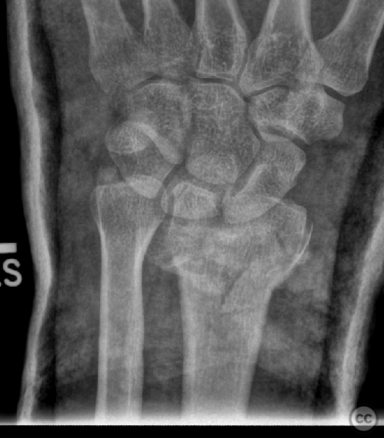
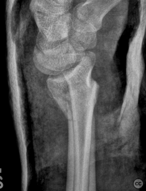
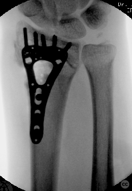
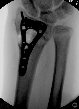
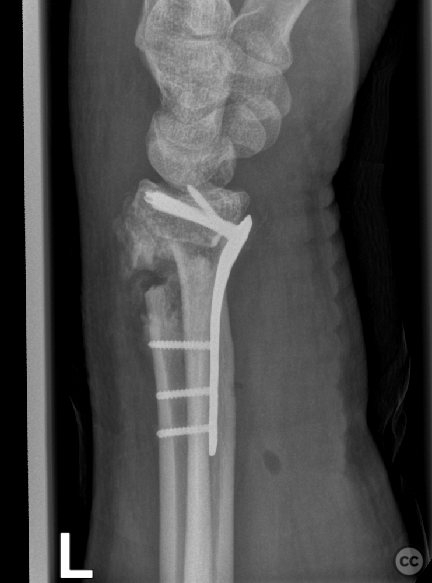
Clinical and radiological findings: 8 week old negelected distal radius fracture
Preoperative Plan
Planning remarks: ostetomy and joint geometry correction
Surgical Discussion
Patient positioning: Supine
Anatomical surgical approach: Henrys
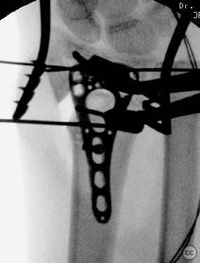
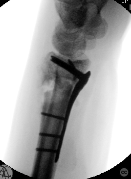
Operative remarks:The patient was positioned in a supine manner on the X-ray translucent operating table, with the left arm placed laterally on an auxiliary arm table. A tourniquet was applied at the proximal end of the ipsilateral arm, followed by sterile preparation and draping of the surgical field. A team time-out was conducted, and two grams of Cefazolin were administered intravenously as prophylactic antibiotics. A longitudinal incision was made over the tendon of the musculus flexor carpi radialis, which was followed by subcutaneous dissection and hemostasis. The tendon sheath of the musculus flexor carpi radialis was incised, allowing for retraction of the tendon toward the ulnar side. The incision of the antebrachial fascia, located above the tendon of the musculus flexor pollicis longus, was also performed, and this tendon was similarly retracted toward the ulnar side, revealing the musculus pronator quadratus at the base of the wound. The incision was extended along the radial border of the musculus pronator quadratus and then along the watershed line, followed by subperiosteal elevation. The distal radial metaphysis was fully visualized. Due to the age of the fracture, approximately seven weeks post-injury, advanced fracture healing was evident. There was no discernible fracture gap at the base of the wound, and no freely mobile fracture fragments were identifiable either in situ or under multiplanar fluoroscopy. Thus, the estimation of the fracture plane was performed under lateral fluoroscopy, followed by the introduction of a 14-millimeter osteotome along the anticipated fracture trajectory from a volar to a dorsal orientation. This maneuver facilitated the restoration of the approximate fracture gap and the mobilization of the distal articular fragment under multiplanar fluoroscopy, utilizing a K-wire distractor at the radial styloid, along with several provisional K-wires. The restoration of joint anatomy was achieved, particularly with regard to radial inclination, translation, volar tilt, and radial length, all of which were restored to physiological limits. Subsequently, a two-column distal radial plate was applied to the distal fragment. The technique involved the insertion of four 2.4-millimeter subchondral screws, which were placed in the distal fragment before the plate was reduced to the distal radial diaphysis, thereby improving the remaining volar radial tilt. The precise positioning of the plate on the distal radial diaphysis allowed for further accurate correction of the distal radioulnar joint. Prior to the placement of a bicortical 2.4-millimeter screw, multiplanar fluoroscopy confirmed the restoration of joint geometry and axis. This was followed by the final placement of two additional 2.4-millimeter bicortical screws to complete the osteosynthesis. The wound was irrigated using a physiological saline solution, followed by the adjustment of the musculus pronator quadratus over the distal plate. The subcutaneous closure was then performed with a running Donati suture to the skin. Finally, sterile dressings and an elastic bandage were applied.
Search for Related Literature

Dr Ed Oates
- Germany , Schleswig Holstein
- Area of Specialty - General Trauma
- Position - Specialist Consultant

Industry Sponsership
contact us for advertising opportunities

Article viewed 614 times
22 Aug 2024
Add to Bookmarks
Full Citation
Cite this article:
Oates, E.J. (2024). Neglected distal radius fracture. Journal of Orthopaedic Surgery and Traumatology. Case Report 6266179 Published Online Aug 22 2024.